📄 New in Neurobiology of Disease. Below, the short story of what it is and why it matters.
The pathway at the heart of Parkinson's disease
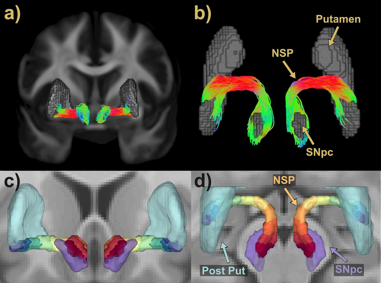
Parkinson's disease involves loss of dopamine neurons that connect the substantia nigra to the striatum through the nigrostriatal pathway.
What we could - and couldn't - see
Molecular imaging shows striatal dopamine loss, but the axonal tract itself is hard to study in vivo.
A brief history of the study
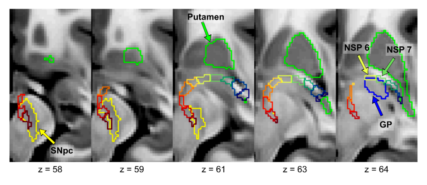
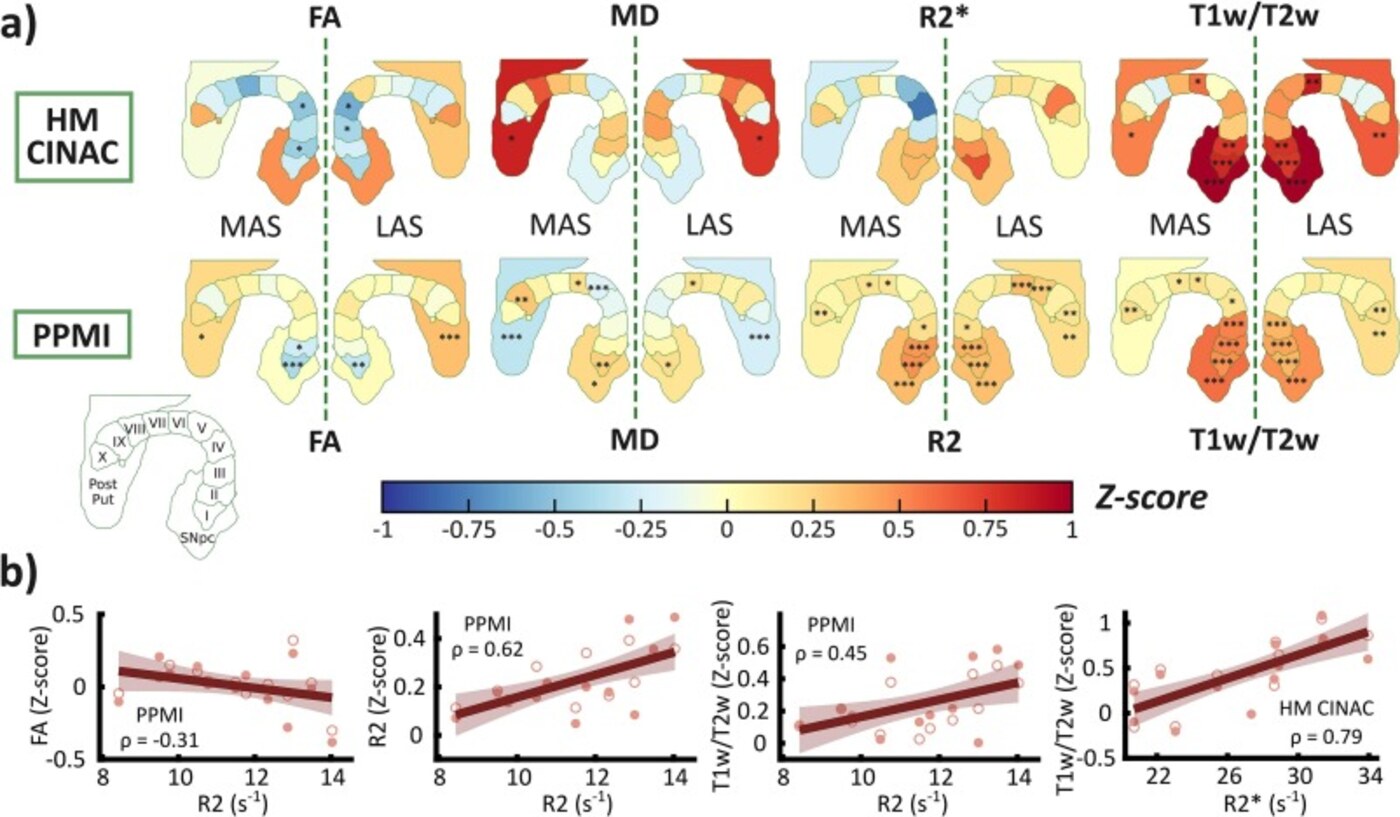
From Miguel López Aguirre's PhD in our lab: we reconstructed the pathway with diffusion MRI and tractography, then quantified its microstructure.
What we found
In early Parkinson's disease, the tract's microstructural changes follow the caudo-rostral pattern of dopamine loss - assessable non-invasively with MRI.
Why it matters
A non-invasive MRI marker of pathway degeneration could help diagnose, stage and monitor Parkinson's disease with widely available imaging.
Congratulations to Miguel and all co-authors - core to our Advanced Neuroimaging field.
Source: López Aguirre et al., Neurobiology of Disease (2026).